Bioimaging refers to the different techniques and systems used for in vivo, ex vivo, and in vitro imaging of biological samples, which allow researchers to obtain information at the molecular, cellular, or tissue level.
Microscopes are widely used by researchers for bioimaging to study subcellular structures, proteins, or organelles. For example, these images help us understand how cells and tissues are organized at the molecular level, identify and study substances in cells such as peptides or RNA, screen for potential drug candidates for the pharmaceutical industry, or serve as a real-time tool for intraoperative surgical margin assessment.
Microscope categories: brightfield and fluorescence
Brightfield microscopy is one of the oldest and most widely used techniques, thanks to its simplicity and easy handling. However, its use of white light can affect the resolution of images obtained (due to the effects of absorption, scattering, or deflection in the sample) and, because some biological specimens are thin and transparent, it can be difficult to achieve detailed, high- contrast brightfield images.
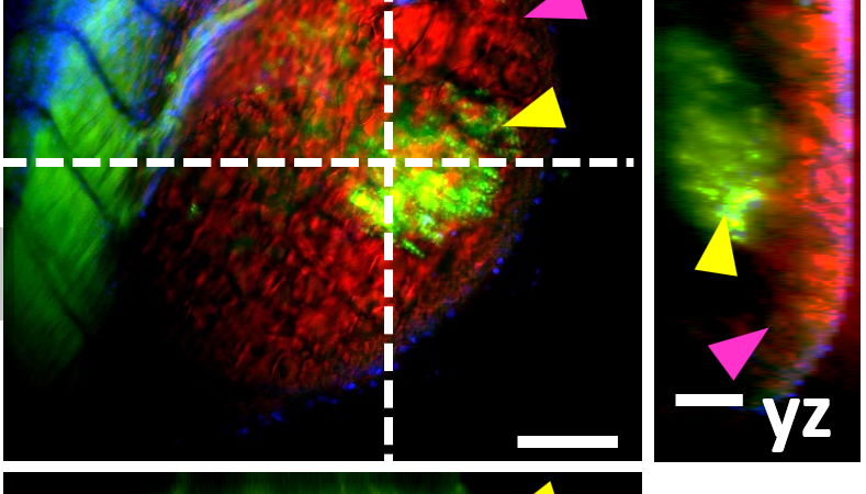
Fluorescence microscopy, which includes fluorescent widefield, confocal, two-photon, or light-sheet microscopy, uses fluorescent stains or proteins called “fluorophores,” which are typically introduced by a researcher. These molecules absorb a specific wavelength of light—the excitation wavelength—and emit a second, longer “emission wavelength.” This technique offers advantages, such as the possibility to detect different molecules simultaneously or to detect them even if not many are present—and single molecules can be imaged.
Read the full article published in Laser Focus World Magazine September 2024 Issue.